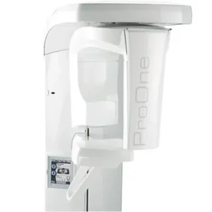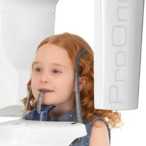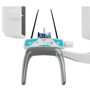


Durr VistaVox S
VistaVox S: a synergy of diagnostic reliability, efficiency, and minimized radiation exposure
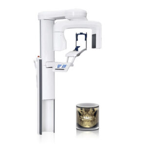
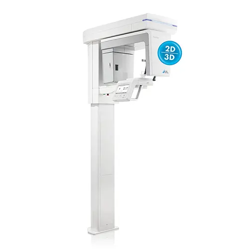
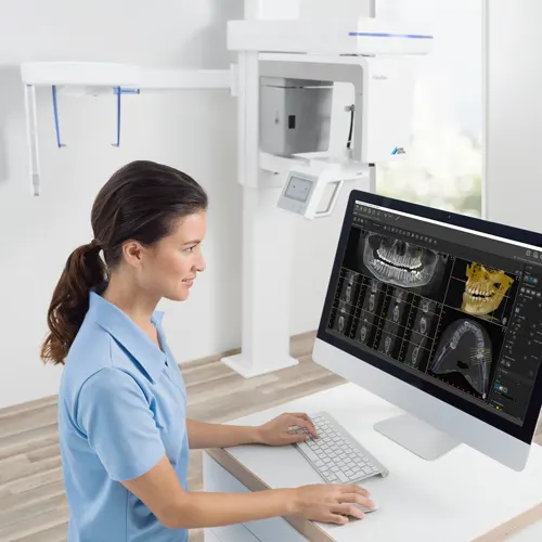
With its ideal imaging volume, effortless positioning, and exceptional image quality, the VistaVox S marks a significant advancement in 3D X-ray systems.
Leveraging its innovative technology, this system produces comprehensive 3D images that facilitate accurate diagnoses, informed treatment decisions, and effective patient communication.
The S-Pan technology of the VistaVox S delivers precisely detailed OPG images, all in the outstanding quality expected from DÜRR DENTAL
VistaVox S offers an ideal 3D volume that is adapted to the jaw arch. The special feature of VistaVox S is that its imaging volume is based on the human anatomy, representing precisely the area you need for diagnostics in the dental region. See what you want to see.
The standout aspect of VistaVox S is its imaging volume, meticulously designed around human anatomy to accurately encompass the specific area essential for dental diagnostics.
VistaSoft 3D
VistaSoft 3D provides diverse visualization options for analyzing CBCT images, including panoramic views, transversal layer images (TSA), and multiplanar reconstruction (MPR). Moreover, the VistaVox system comes with VistaSoft as an unlimited network license, along with a floating license that supports up to three 3D workstations.
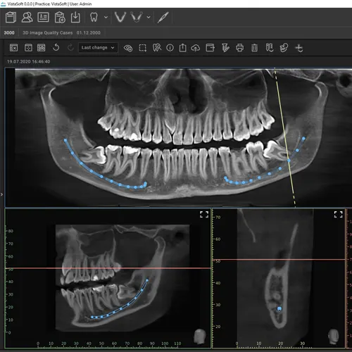
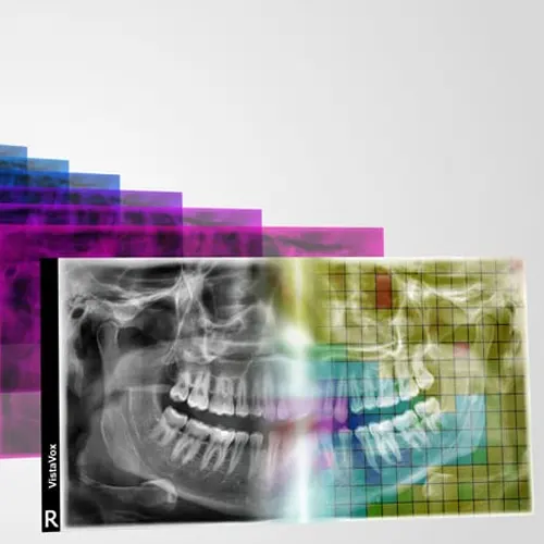
S-Pan technology improves diagnostic reliability with sharp images.
Utilizing S-Pan technology, the regions of an image that most accurately reflect the patient’s anatomy are seamlessly selected from a multitude of parallel layers.
The outcome is a remarkably clear image, allowing for quick and easy identification of all anatomically significant structures.
Furthermore, the reconstruction is precisely aligned with the patient’s bite position, effectively compensating for any misalignment.
This not only streamlines the surgical process but also spares the patient from undergoing repetitive imaging.

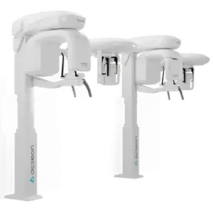
VistaVox S Ceph: The Ultimate 3-in-1 X-Ray Solution
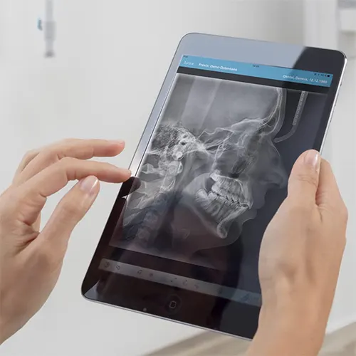
VistaVox S Ceph not only includes a diverse range of CBCT volumes and 17 panoramic programs, but it also features six distinct modes tailored for all cephalometric exposure types.
With an incredibly swift scan time starting at just 1.9 seconds, it effectively minimizes motion artifacts and reduces radiation exposure.
Additionally, the state-of-the-art high-sensitivity CsI sensors ensure outstanding image quality.

Reduction of 3D Metal Artifacts (MAR)
Metal artifacts pose a significant challenge for 3D imaging. During three-dimensional reconstruction, radiopaque objects create shadows—areas that appear completely white—and streaks.
This is especially problematic for patients with metal prosthetics, complicating diagnostic efforts.
However, VistaVox MAR effectively eliminates these metal artifacts using cutting-edge algorithms, resulting in a far clearer representation of anatomical structures.
| VistaVox S | VistaVox S Ceph | |
|---|---|---|
| Image detector type | CsI CMOS Photodiode array | Csl CMOS Photodiode array |
| Pixel size (µm) | 49,5 | 49,5 / 100 |
| Scan times (s) | 2 – 18 | 1,9 – 18 |
| Active sensor surface area (mm) | 135,8 x 36,4 | 135,8 x 36,4 / 157,2 x 16,3 |
| Voltage (kV) | 60 – 99 | 60 – 99 |
| Tube current (mA) | 4 – 16 | 4 – 16 |
| Focal spot (mm) | 0,5 | 0,5 |
| Min. total filtration (mm) | 2,8 | 2,8 |
| 3D volume, child, diagnostic (mm) | Ø 130 x 70 | Ø 130 x 70 |
| 3D volume, adult, diagnostic (mm) | Ø 130 x 85 | Ø 130 x 85 |
| 3D volume, jaw region (mm) | Ø 50 x 50 | Ø 50 x 50 |
| Height (mm) | 1.406 – 2.250 | 1.406 – 2.206 |
| Weight (kg) | 180 | 202 |
| Height adjustment range (mm) | 844 | 800 |
| Width x depth (mm) | 990 x 1.130 | 1.941 x 1.615 |
| Installation (wall-mounted) | ||
| Frequency (Hz) | 50/60 | 50/60 |
| Rated power (VA) | 2.200 | 2.200 |
| Mains voltage (V AC) | 200 – 240 | 200 – 240 |